
Artificial intelligence (AI) is one of the most powerful tools in the history of medicine, capable of accelerating diagnoses, personalizing treatment, and transforming care at a scale never before possible. But most advances so far have been built for adults.
Սթենֆորդի բժշկական կենտրոնի մանկական առողջություն is racing to close that gap—and to build a future where every child can benefit from AI’s full potential.
Stanford is launching the Hub for Artificial Intelligence in Pediatric Medicine, dedicated entirely to developing and deploying AI solutions for children’s health. The Hub will ensure that advances in cutting-edge research and technology can change the standard of care for kids at Stanford Children’s and beyond, in ways that are safe, equitable, and patient-centered.
No institution is better positioned to lead this work. Stanford brings together trailblazing researchers, a world-class children’s hospital, and unmatched computer science expertise, all at the epicenter of the global technology industry. Across specialties, from cancer to congenital heart disease to complications of prematurity, Stanford is already transforming pediatric care.
Until now, these efforts have largely operated in parallel. The Hub will change that, uniting doctors, researchers, engineers, ethicists, and data scientists to enable progress that no single team could achieve alone.
A catalytic $10 million gift from Alfred E. Mann Charities will help recruit a world-class leader to be the connective force, driving this work forward.
The result will be a new era in children’s health, one in which the most powerful tools in medicine can reach the children who need them most, in the Bay Area and around the world.
Ռeinventing MRI for Children
If you’ve had an MRI exam, you know how uncomfortable it can be to lie still in a loud, narrow tube, sometimes for nearly an hour.
Now, imagine how difficult that would be for a young child. Many pediatric patients need to be sedated during an MRI to remain still. But sedation can add cost and complexity, all while increasing medical risks.
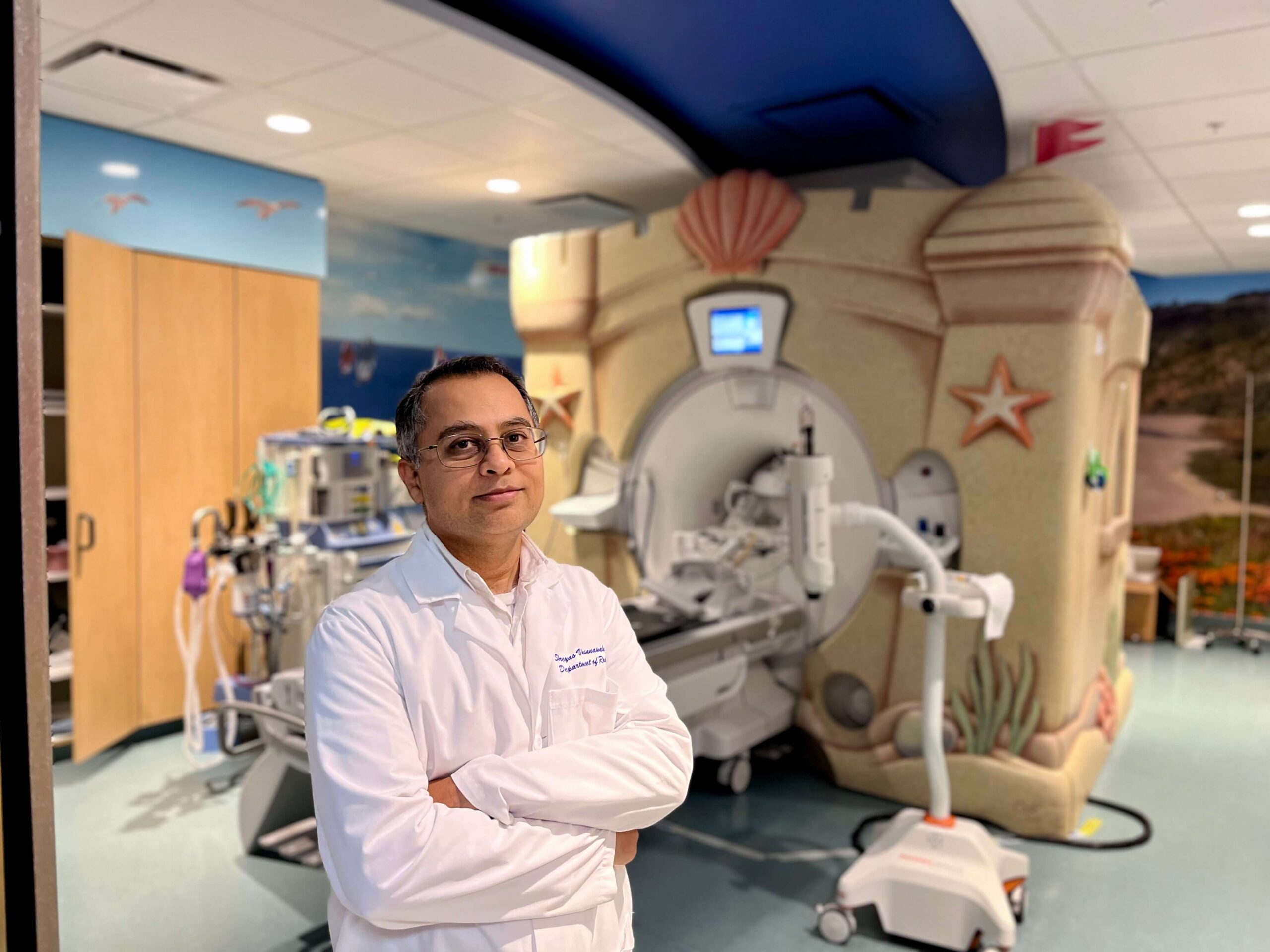
Shreyas Vasanawala, MD, PhD (shown above), and collaborators across Stanford and the Bay Area are tackling this challenge by pioneering the use of AI to make pediatric MRI scans faster, less expensive, and even more detailed.
The Vasanawala Lab is using machine learning to make this possible. By training AI on many MRI scans, the system learns what detailed images should look like. It can then take a much shorter, lower-fidelity scan and fill in missing information, reconstructing a clear, complete, and accurate image from limited data. Scanning time can now be just a few minutes instead of an hour.
“The AI algorithms we developed can now recover an unprecedented level of detail,” says Vasanawala. “You can see a tiny artery in an infant that would otherwise be blurred. And in some cases, these details allow us to make a diagnosis that would have been impossible in the past.”
One of these algorithms has already been cleared by the Food and Drug Administration and is improving imaging for kids with congenital heart disease. It is being used daily in more than 160 countries and has already reached more than 80 million patients.
The impact on patients is significant. Shorter scans mean fewer children require anesthesia. And even when sedation is needed, its depth and duration can be reduced.
Faster exams also mean shorter wait times to get appointments for MRIs, which have historically taken months in some locations. AI-enhanced scans also empower MRI technologists to tackle more complex exams, meaning that more patients can have access to better-quality scans, even in countries with fewer resources.
Meanwhile, Vasanawala’s lab is continuing to drive innovation. His team’s ultimate goal is to automate the entire imaging process, from identifying the type of scan needed and precisely locating the area of concern to performing the scan and correcting image distortions in real time—all within minutes.
“What if getting an MRI were as simple as measuring blood pressure?” says Vasanawala. “That’s the promise of AI: empowering doctors with tools to see more clearly, act more quickly, and ultimately change lives.”
A New Era in Care: Three AI breakthroughs rewriting the odds for moms and kids
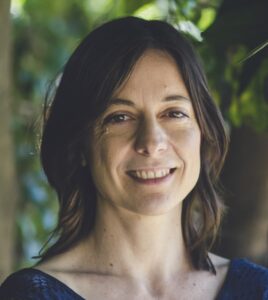
Spotting Risk Early—Ivana Maric, PhD
Preeclampsia—dangerously high blood pressure during pregnancy—impacts about 8% of expectant mothers worldwide. With roughly 10 million cases each year, it is a leading cause of maternal death and preterm birth, creating lifelong health complications for millions.
There is a desperate need to identify preeclampsia risk early, precisely, and affordably, and to develop effective interventions that safeguard the health of both mother and baby.
Ivana Maric, PhD, and her team are developing a simple, low-cost test to detect preeclampsia risk long before symptoms appear. Her team used machine learning to study hundreds of biological markers in the blood. Then, they identified a specific protein ratio that can be measured with a simple urine test as early as 10 to 12 weeks into pregnancy, well before symptoms arise and in time for preventive therapies like a low-dose aspirin regimen to be most effective.
The test is now being validated in two larger studies—one of them international. Once that is completed, the test could make early, personalized prenatal screening available to women everywhere, regardless of where they live or what resources are available to them.
“If we can predict early in pregnancy who is at risk of preeclampsia, we can provide those moms with a preventive treatment,” says Maric. “This could help prevent tragic outcomes for mothers and the complications of preterm births for babies.”
Protecting Preemies—Nima Aghaeepour, PhD
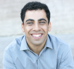
Premature babies can develop life-threatening complications in their first months of life, often without warning. Nima Aghaeepour, PhD, a world leader in applying AI to maternal and newborn health, is working to change that.
In earlier work, Aghaeepour and his Stanford collaborators developed a way to use AI to personalize IV nutrition for fragile preemies—reducing medical errors and improving care in low-resource settings. Now he is focusing on predicting and working to prevent the most serious complications preemies can face.
His team analyzed routine blood samples from more than 13,000 very premature babies, along with their medical records. They looked at which infants went on to develop one of four major types of complications linked to prematurity, such as bronchopulmonary dysplasia, a lung disease, or bleeding in the brain.
In their study, published in Science Translational Medicine, they used AI to identify patterns in blood molecules that were correlated with an infant later developing a major complication.
Based on this information, the team built a tool that could predict these complications with 85% certainty. In the future, this tool could help doctors identify which babies are most at risk and allow them to intervene earlier to prevent life-threatening health issues.
“It’s a complete change in the way we think about prematurity,” says David Stevenson, MD, director of the Prematurity Research Center at Stanford.
Expanding Diabetes Care—David Maahs, MD, PhD, and Priya Prahalad, MD, PhD
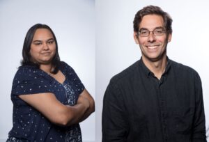
Managing diabetes means keeping blood sugar levels in a healthy range. But for care teams, keeping tabs on multiple patients is time consuming. Led by David Maahs, MD, PhD, and Priya Prahalad, MD, PhD, researchers used AI tools to create a smart dashboard to improve care for teens with type 1 diabetes.
This dashboard gathers and filters multiple data points from many patients at once. It shows who is consistently wearing their continuous glucose monitors and keeping their blood sugar in the target range. That allows the care team to focus on patients who need extra help—for example, with getting a new glucose monitor prescription or adjusting insulin doses.
The results are impressive. Maahs and Prahalad’s study found that after the dashboard was introduced, the number of patients keeping their blood sugar at healthy levels more than doubled. Importantly, patients from all backgrounds benefited equally, regardless of their insurance status.
“Often, when new medical technology becomes available, some patients are left behind,” says Maahs. “It is very encouraging that patients from all backgrounds benefited from the program.”
Բարեգործության կողմից աջակցվող
To learn more about scientific advances in this field at Stanford and philanthropic opportunities to support the AI Hub, contact Charlie.White@LPFCH.org.