
Everything about Elizabeth Rodriguez-Garcia’s pregnancy had been perfectly normal. She’d had morning sickness on occasion and felt a little tired some days, but at 25 weeks, the only real news she and her husband, Salvador Alvarez, expected to hear during a routine ultrasound in July 2013 was the sex of their baby. In anticipation, the couple even brought his mother and her grandmother along to the appointment at her local obstetrician’s office in Salinas so they could see the baby’s images, too.
As the family crowded into the room and the lights were dimmed, the first grainy black-and-white pictures appeared on the monitor. Everyone was excited to see the baby’s tiny body, head, arms, and legs. Within minutes, however, it was clear something was wrong.
The ultrasound technician found an unexpectedly large dark spot where the baby’s left lung should be. A more detailed image was needed and Elizabeth was sent five miles across town to the Stanford Children’s Health Perinatal Diagnostic Center in Salinas, where technicians conducted another scan.
As a nervous Elizabeth waited for news about what might be wrong, her ultrasound images and medical records were being digitally sent 80 miles to the Center for Fetal and Maternal Health at Lucile Packard Children’s Hospital Stanford.
In an instant, both mother and baby became a concern of the center’s medical director, Susan Hintz, MD, MS Epi, and a team of doctors who would address every aspect of their care—from the simple to the complex, and everything in between.
Established in 2011, the Center for Fetal and Maternal Health, part of our hospital’s Johnson Center for Pregnancy and Newborn Services, provides comprehensive care and critical services to mothers and babies in high-risk pregnancies throughout the Bay Area and beyond.
Jumping into Action
Almost as soon as Elizabeth’s ultrasound arrived at our hospital, Hintz and several pediatric and obstetric specialists began examining her images and medical records. Stephanie Neves, administrative coordinator for the center, got to work setting up an array of appointments. By the time Elizabeth and Salvador stepped inside the hospital a few days later, Hintz and her team was ready to give them a diagnosis, a prognosis, and a plan that they believed could save the baby’s life.
“Even before all of us met with Elizabeth face to face, there had been many, many meetings to review the ultrasounds, the literature, and our experience, and to formulate a plan,’’ says Hintz. “We wanted to offer her the best and safest approach.”
The diagnosis was rare: a congenital pulmonary airway malformation, also known as a CPAM. The baby had developed a large, abnormal cyst in the lower left side of his lung. The cyst, full of fluid, was impeding growth of the lung; it was so large that it also was compressing his esophagus and pushing on his heart.
Causing even more concern, a new ultrasound conducted that morning showed that in just a few days the cyst had grown even larger. The baby was collecting more fluid than expected and was at risk of dying in utero from a condition known as hydrops.
After much consultation, the doctors told Elizabeth and Salvador that inserting a shunt through her to the baby and draining the cyst into the amniotic fluid offered the baby the best chance of survival.
Elizabeth and Salvador agreed. A week after the cyst was first found, Jane Chueh, MD, director of prenatal diagnosis and therapy, inserted a large needle into Elizabeth’s abdomen, led it through the baby’s chest, and placed a small rubber shunt through the needle into the cyst. It was the first time Chueh had performed this procedure at Lucile Packard Children’s Hospital Stanford.
“It immediately started to drain,’’ says Chueh. “It was like popping a water balloon. All of the fluid came out in seconds.”
Relieving pressure from the cyst came at a critical time, adds Chueh. Although the cyst partially refilled the following day, it stabilized at a more manageable size for the baby and didn’t recreate the earlier pressure on his chest. More importantly, the fluid retention or hydrops that doctors worried was endangering the baby’s life improved dramatically.
Watching and Planning
After the procedure, Elizabeth initially lost then quickly regained her amniotic fluid and spent a few more weeks undergoing near-daily ultrasounds to make sure the baby was okay. At 30 weeks, Elizabeth was ready to be discharged. But Hintz and her team worried that a complication might develop or that Elizabeth could go into preterm labor, so rather than send her back to Salinas—90 minutes away—a social worker provided her with a fully furnished apartment just minutes down the road, where she could be close by if something unexpected happened.
In the meantime, Hintz and her colleagues drew up a new plan—this one for delivery. A fetal MRI, taken at 37 weeks, helped assess the amount of normal lung tissue and showed that once the baby was born and relying on his own lungs to keep him alive, the cyst would need to be removed immediately to prevent his breathing from being obstructed.
Importantly, notes radiologist-in-chief Richard Barth, MD, the fetal MRI spared the baby from having to undergo a CT scan to further delineate the lung mass—involving radiation and possibly additional anesthesia exposure—after birth and prior to undergoing the much-needed surgery.
Elizabeth carried her baby to 39 weeks of pregnancy, much longer than anticipated.
To simplify the transition between procedures, a Cesarean section was scheduled for an operating room, instead of in the labor and delivery unit, which allowed the baby to be quickly moved to a fully staffed and prepped operating room right after birth.
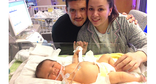
On November 25, 2013, a team of three dozen medical specialists assembled in two adjoining rooms. There were neonatologists and anesthesiologists. Radiologists and surgeons. Nurses and respiratory therapists. Hintz was there, as was Chueh, who delivered the baby by C-section, and Alexis Davis, MD, a neonatologist, who had participated in every discussion about the baby’s care and health.
“We had everyone on deck,’’ says Davis. “We had to be prepared because we knew he could have significant lung and breathing problems at birth.”
Standing by and waiting just for the baby, who would be named Elijah, was a team of doctors and medical experts led by pediatric surgeons Karl Sylvester, MD, and Matias Bruzoni, MD. Within minutes of his birth, Elijah was moved quickly into the nearby operating room. In a two-hour procedure, Sylvester removed both the cyst and more than two-thirds of the baby’s lung that was adversely affected by the growth.
“Our ability as an institution to provide all these subspecialists in two rooms to care for both the mom and the baby is what led to the successful outcome for this family,’’ Sylvester says. “It made a huge difference in this young family’s life; without it, he may not have survived at all.”
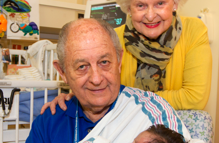
Elijah remained in the hospital’s Neonatal Intensive Care Unit, where he was closely monitored for a month, while his parents stayed nearby at the Ronald McDonald House at Stanford.
On Christmas Eve, Elijah’s parents took him home to Salinas—the best gift they could ever have, says Salvador.
Hope for the Future
It’s too soon to tell what the longer term effects will be on Elijah’s lung, says Sylvester. Lungs continue to grow and remodel until a child is about 7 years old, so there is a good chance Elijah’s lungs will grow to a normal size. His path so far is encouraging and his doctors continue to manage and monitor his care.
“This was a fantastic outcome for Elijah and his family,’’ says Hintz. “Our multidisciplinary team carefully and thoughtfully considers the best treatment approach for each of these challenging cases. We are extremely fortunate to have the expertise and experience here at Lucile Packard Children’s Hospital to assure the best possible outcomes for extremely complex fetal patients and their families.”
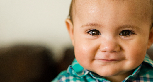
“He looks like a completely normal baby,’’ Elizabeth says. “If you see him, you’d never know what he went through and that he doesn’t have most of his left lung. The cyst is completely gone.”
Salvador describes Elijah as a healthy, active, and happy baby, whose only physical sign of his near-catastrophic condition is the fading seven-inch scar on his chest. He has a big appetite and loves to laugh and play. He pulls himself up and walks along the family’s furniture.
“I feel blessed to have met the doctors at Lucile Packard Children’s Hospital,” adds Elizabeth, who recently returned to work as a nursing assistant. “I felt safe and comfortable the whole time I was there. I felt like they were my family and that I could trust them.”
This article first appeared in the Fall 2014 issue of Lucile Packard Children's News magazine.