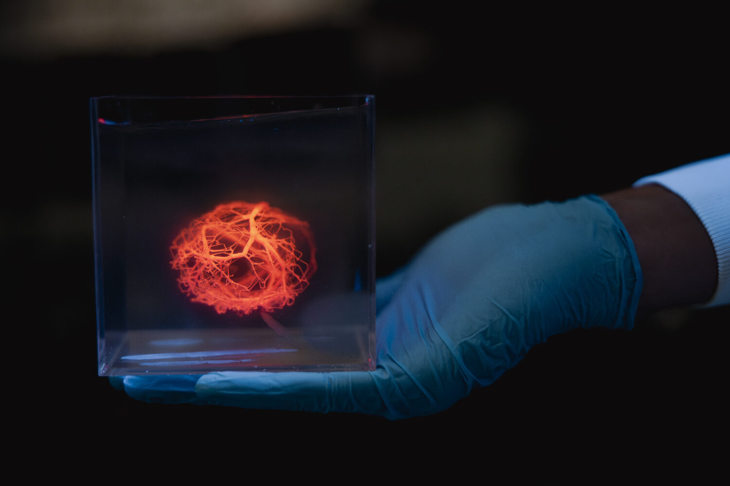
A model of a vascular tree printed using a 3D bioprinter. | Andrew Brodhead
Stanford researchers have taken an exciting leap forward in their quest to 3D print a human heart, according to a new paper published in the journal Science.
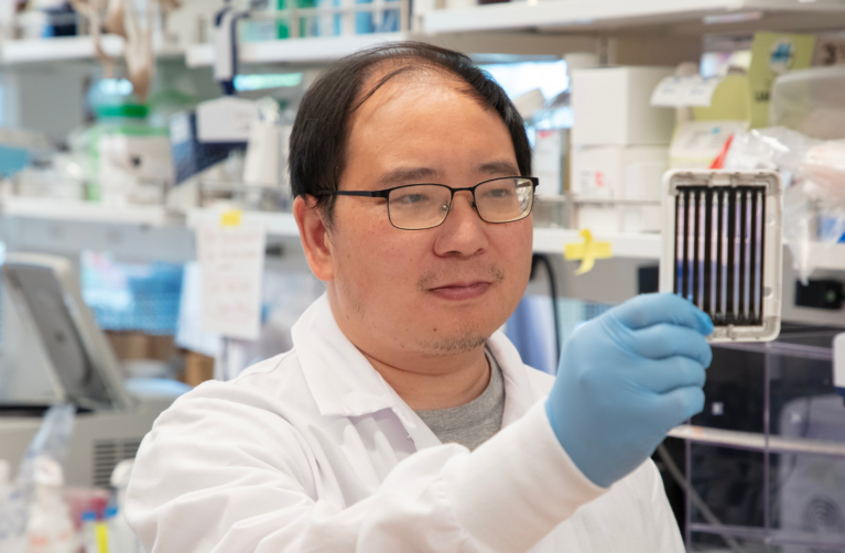
On this mission are Alison Marsden, a professor of pediatrics and bioengineering, and Mark Skylar-Scott, an assistant professor of bioengineering and a member of Stanford’s Basic Science and Engineering (BASE) Initiative, which aims to transform the future of care for kids with congenital heart disease. In the paper, the senior co-authors announced new tools to design and 3D print the incredibly complex networks of functioning blood vessels (i.e., “vascular trees”) needed to carry blood throughout a 3D printed organ.
Help bring revolutionary solutions to kids with congenital heart disease.
Marsden and her colleagues—using advanced computer modeling and simulation tools—developed an algorithm that can create vascular trees that closely mimic real ones, and they’ve made the software available for anyone to use. It’s 200x faster than prior methods. In the lab, they are now able to generate a computer model of a vascular tree in five hours versus the several months it would’ve taken with previous algorithms.
The researchers are quick to note that this is a great first step, but much more work needs to be done to build fully functional blood vessels. Skylar-Scott calls it a “critical step in the process” of bioprinting an entire human heart using a patient’s own stem cells. This research has great potential to help children with heart disease, as well as children who are on an organ transplant waitlist or those who have experienced organ rejection.
Philanthropy is essential to the BASE program—helping to recruit a dream team of basic scientists and engineers, and funding trailblazing research into pediatric heart disease.